
chemistry
47th Annual Naff Symposium
|
Innovation in Molecular Neuroscience Schedule of Events - April 1, 2022 |
|
|---|---|
| 8:00am |
Registration and Continental Breakfast |
| 8:50am |
Welcome - TBD |
| 9:00am |
Dr. Erin Calipari |
| 10:00am |
Break |
| 10:30am |
Dr. Tim Harris |
| 11:30am |
Lunch & Break |
| 1:00pm |
Dr. Elizabeth Hillman |
| 2:00pm |
Break & Poster Session Set-Up |
| 2:30pm |
Dr. Baljit Khakh |
| 3:30 - 5:00pm |
Poster Session |
|
Speakers |
|
|---|---|
 |
Dr. Erin Calipari Vanderbilt University Dr. Calipari received her PhD in Neuroscience in 2013 in the laboratory of Dr. Sara Jones at Wake Forest University School of Medicine where she studied how self-administered drugs altered dopaminergic function to drive addictive behaviors. She then went on to complete her postdoctoral training with Dr. Eric Nestler at Icahn School of Medicine at Mount Sinai, where she used circuit probing techniques to understand the temporally specific neural signals that underlie motivation and reward learning. She is currently an Assistant Professor at Vanderbilt University in the Department of Pharmacology. Her independent work seeks to characterize and modulate the precise circuits in the brain that underlie both adaptive and maladaptive processes in reward, motivation, and associative learning. |
 |
Dr. Tim Harris Johns Hopkins University Timothy Harris is a research professor in the Department of Biomedical Engineering. He leads the Applied Physics and Instrumentation Group at the HHMI Janelia Research Campus, and is the originator of the project that produced the Neuropixels Si probe for extracellular recording in animals, mostly mice, and rats. He shares his time between Janelia and Johns Hopkins and is working on projects to enable recording 10-20,000 neurons in rodents and 30-50,000 neurons in non-human primates, as well as stimulate with high resolution. He received a BS in Chemistry at California Polytechnical State University, San Luis Obispo, and a PhD in Analytical Chemistry at Purdue University. |
 |
Dr. Elizabeth Hillman Columbia University Elizabeth Hillman is professor of biomedical engineering and radiology at Columbia University and a member of the Mortimer B. Zuckerman Mind Brain Behavior Institute and Kavli Institute for Brain Science at Columbia. Hillman received her undergraduate degree in physics and Ph.D. in medical physics and bioengineering at University College London and completed post-doctoral training at Massachusetts General Hospital/Harvard Medical School. In 2006, Hillman moved to Columbia University, founding the Laboratory for Functional Optical Imaging. Hillman’s research program focuses on the development and application of optical imaging and microscopy technologies to capture functional dynamics in the living brain. Most recently, she developed swept confocally aligned planar excitation (SCAPE) microscopy, a technique capable of very high speed volumetric imaging of neural activity in behaving organisms such as adult and larval Drosophila, zebrafish, C. elegans and the rodent brain. Hillman’s research program also includes exploring the interrelation between neural activity and blood flow in the brain, as the basis for signals detected by functional magnetic resonance imaging (fMRI). Hillman is a fellow of the Optical Society of America (OSA), the society of photo-optical instrumentation (SPIE) and the American Institute for Medical and Biological Engineering (AIMBE). She has received the OSA Adolf Lomb Medal for contributions to optics, as well as early career awards from the Wallace Coulter Foundation, National Science Foundation and Human Frontier Science Program. |
 |
Dr. Baljit Khakh University of California, Los Angeles Baljit Khakh completed his Ph.D. at the University of Cambridge in the laboratory of Patrick PA Humphrey. He completed postdoctoral fellowships in the laboratory of Graeme Henderson at the University of Bristol, and then in the laboratory of Henry A. Lester and Norman Davidson at California Institute of Technology. In 2001, Khakh became Group Leader at the MRC Laboratory of Molecular Biology in Cambridge, and in 2006 he moved to the University of California, Los Angeles where he is Professor of Physiology and Neurobiology. Khakh’s work has been recognized, including with the NIH Director's Pioneer Award, the Paul G. Allen Distinguished Investigator Award, and the Outstanding Investigator Award (R35) from NINDS. |
2022 Naff Symposium Committee
Dr. Chris Richards - Chair
Jason DeRouchey (Chemistry)
Lance Johnson (Physiology)
Brandon Henderson (Marshall University)
Date:
-
Location:
WT Young Library Auditorium
Tags/Keywords:
Event Series:
Jack Steele (Ph.D. 1968) - A Biography
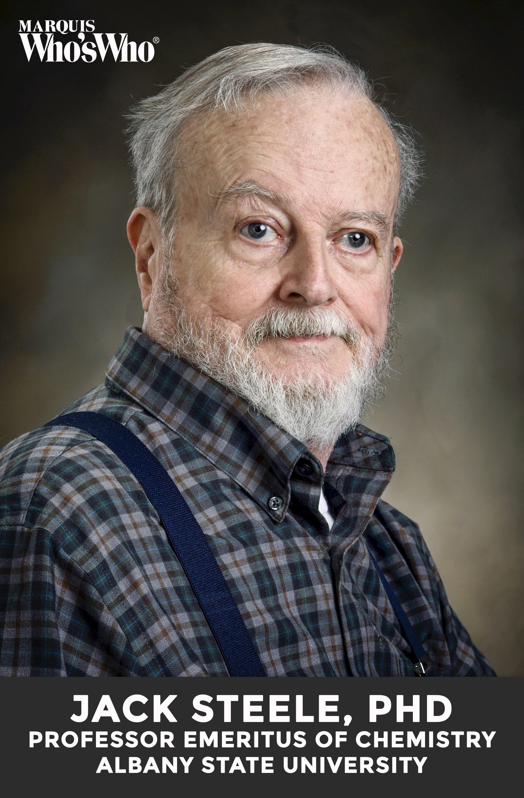 As a 6th grade student in his hometown of Greencastle, Indiana, Jack Steele realized that his life ambition was to be a chemist and, when time came to go to college, he pursued a BA in chemistry at DePauw University. Jack worked on electrochemistry with Prof. Eugene Schwartz at DePauw the summer of 1964 after getting his BA.
Obituary: Paul G. Sears
This article previously appeared in Chemical and Engineering News on November 16.
Paul G. Sears, 96, died September 12 in Lexington, KY.
Glazer and Heidary Award from the National Science Foundation
The National Science Foundation has awarded a new grant to Drs. David Heidary and Edith Glazer for the development of chemical tools to study RNA. The project, titled “Inorganic-aptamer hybrids for live cell imaging”, leverages the complementary expertise of the investigators in the development of optical cellular assays and the creation of photoactive inorganic molecules.
Charles Herschel Holmes Griffith: Laboratory Supervisor & Legend
By J. Susan Griffith, M.D.